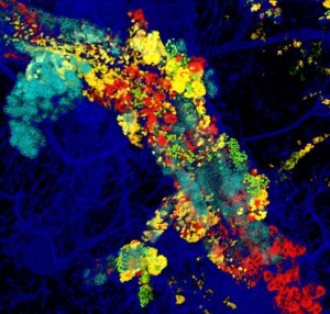

Image supplied by Dr Anne Rios. This image, taken using the new single-cell resolution 3D imaging technique, shows a mammary duct with many precancerous clones (‘sister’ cells of one colour) within the tissue. Only a small proportion of these precancerous cells will develop into tumours.
The image looks like beautiful modern art, but they are so much more than that.
For the first time, scientists from the Walter and Eliza Hall Institute of Medical Research (WEHI) in Melbourne have published images of breast cancer taken using a new visualisation technique, called large-scale single-cell resolution 3D imaging. These highly-colourful and unique images will help researchers learn more about how cancer cells interact with each other. With further study, the state-of-the-art technique may be applied to obtain more comprehensive information about molecular markers and tumour architecture that can aid clinical diagnosis of breast cancer.
Most routine imaging techniques used by pathologists to diagnose breast cancer alter or destroy the tissue structure. Remarkably, the new technique developed by Dr Anne Rios, who was funded by NBCF, does not. This allows scientists to view the cancer cells within a biopsied sample in a more natural setting, so that they can see how the cells arrange themselves and interact with each other. Dr Rios has used her new imaging technique in both normal human breast tissue and a number of patient-derived breast cancer tumours. The resulting colourful images were recently published in the prestigious journal Cancer Cell.
In short, the technique enables 3D viewing of the tissue at an incredibly high resolution. The biopsied tumour sample is processed using a single-step, non-toxic agent which allows imaging of the entire piece of tissue with unprecedented clarity, down to every cell that constitutes the breast (nanometre scale). The researchers are then able to combine multiple images into videos, such as here, to view tumours in a completely new way.
Dr Rios is excited about the possibilities this new imaging technology may open for women with breast cancer. “This technique will hopefully become the gold standard for imaging breast cancer samples,” she said.
This work was completed at the WEHI, where Dr Rios was a postdoctoral fellow under the supervision of Professor Geoffrey Lindeman and Professor Jane Visvader. Dr Rios now heads her own laboratory and the Imaging Centre at the Princess Maxima Centre for Pediatric Oncology in the Netherlands. NBCF is proud to have contributed to the funding of this study.
More News Articles
View all News


