Surgical removal of breast cancer tumours is an effective treatment method, but it can be challenging to remove all the cancer cells. If cells remain, then they can grow and spread, leading to a recurrence of cancer. Currently, approximately one in four women will require additional surgery to remove these missed cells.
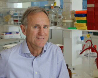
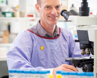
A new quantitative imaging method from Associate Professor Brendan Kennedy (Harry Perkins Institute of Medical Research & The University of Western Australia) may improve these odds. Until recently, surgeons were highly reliant on their vision and touch to determine whether the entire tumour had been removed. X-ray imaging could also be used, but this technique cannot detect microscopic traces of tumour that can be left behind.
“Despite living in the ‘digital age,’ surgeons must routinely rely on their eyesight and sense of touch to determine if they have removed the entire tumour during breast-conserving surgery,” said A/Prof Kennedy. “Due to lack of adequate tools, 20 to 30 percent of patients must return for additional surgery, resulting in substantial physical and financial burdens and increased risk of complications.”
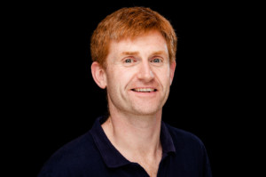
Associate Professor Brendan Kennedy
The new technique, published recently in the journal Cancer Research, utilises a high-resolution three-dimensional imaging method called optical coherence tomography (OCT).
“OCT can be described as the optical equivalent of ultrasound, using reflections of light waves rather than sound waves to form images of tissue microstructure,” A/Prof Kennedy explained.
In this case, a specialised form of OCT imaging called quantitative micro-elastography (QME) has been adopted to provide three-dimensional maps of tissue elasticity at the margins of the tumour. Cancerous tissue is stiffer than normal tissue, which allows QME to detect areas which should be removed by the surgeon.
In the study, researchers took surgical samples from 90 women undergoing breast cancer surgery. They then compared the QME technique to standard histology, showing that the new method has over 95% accuracy for detecting cancer within 1mm of the surgical margin.
“The ideal scenario would be to perform the imaging in the surgical cavity immediately after the specimen has been removed,” A/Prof Kennedy said. “This would give surgeons a direct indication of whether any tumour had been missed. As such, our next goal is to develop a handheld QME probe to enable intraoperative imaging.”
“We are grateful to NBCF for providing seed funding in the early stages of developing the technology which was instrumental in enabling us to get to where we are today” A/Prof Kennedy said. This study was supported by OncoRes Medical, Cancer Council Western Australia and the Department of Health Western Australia.
More News Articles
View all News