Sentinel Lymph Nodes Biopsy
Sentinel Lymph Nodes
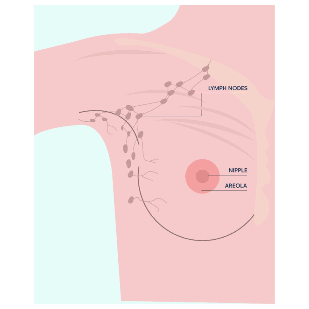
Lymph nodes are part of the lymphatic system, which make up part of the body’s immune system. Lymph nodes are small, rounded masses of lymphatic tissue located along the lymph vessel. They filter lymph fluid to remove harmful substances (such as bacteria, viruses and abnormal cells such as cancer cells) and return the ‘clean’ fluid back to the bloodstream.
As such, they are vital for fighting infections. Lymph nodes are located all over your body, including your underarms, groin, neck, the area around the lungs and the gut.
What are sentinel lymph nodes?
A sentinel lymph node is the first lymph node to where cancer cells may spread outside of the primary tumour site (such as the breast). Generally, the sentinel node is located in the underarm (axilla), however it can also be found in other parts of the body, such as in the chest between the ribs under the breast or above or underneath the collarbone. Typically, people have one sentinel node, but it’s possible to have two or three.
How is a sentinel node biopsy performed?
A sentinel node biopsy involves mapping the location of the node (or nodes) either before or during a biopsy or mastectomy procedure. To help locate the sentinel node, the patient is injected with a small amount of a low-grade radioactive fluid and/or blue dye in the breast around the cancer or under the nipple area. The radioactive fluid flows to the sentinel nodes through the lymphatic vessels and can be seen on a nuclear medicine scan (lymphoscintigram).
Lymphoscintigraphy is used to find the sentinel lymph node (or nodes). The blue dye also flows through the lymphatic vessels to the draining lymph nodes of the cancer.
With the use of a hand-held probe (a highly directional Geiger counter) that detects the radioactive substance together with the blue appearance of the sentinel node, the surgeon is guided to the correct node(s) where a small incision in the skin is made to remove the node (or nodes).
What happens after a sentinel node biopsy?
After the sentinel node biopsy, a pathologist checks the sentinel node for cancerous cells. If cancer is present, the surgeon will remove additional lymph nodes. If no cancer cells are detected, the additional removal of lymph nodes will not be required.
What are the possible side effects of sentinel node biopsy?
Potential side effects of a sentinel node biopsy include:
- There is a low risk of shoulder stiffness, numbness in the arm, shoulder, underarm and parts of the chest; seroma (fluid collecting near the surgical scar); lymphoedema in the arm (built up of lymph fluid) causing swelling, pain or discomfort.
- There is a small risk of skin/allergic reactions to the blue dye test or radioactive fluid.
- The use of the blue dye to find the sentinel node could turn the urine a blue-green colour for 24 hours after surgery. The skin of the breast could also turn blue, but this fades away with time.
- A false-negative biopsy result is possible – the doctor and patient are misled into thinking the cancer cells have not spread to nearby nodes.
Frequently Asked Questions
Potential side effects after a sentinel node biopsy may include arm or shoulder stiffness, numbness in the arm, shoulder, underarm and parts of the chest, seroma and lymphoedema and the severity of these side effects will depend on the number of nodes removed. There is a small risk of a mild allergic reaction to the radioactive fluid or blue dye. See above for more on side effects.
Recovery time following a sentinel node biopsy varies from person to person. Most people will feel close to normal in a few days. The incision usually heals in about two weeks. If a sentinel node biopsy is done at the same time as other breast surgeries (such as lumpectomy or mastectomy) recovery will be different. In contrast lymph node removal could include axillary dissection/axilliary clearance, which is the removal of several or all the lymph nodes under the armpit. These leads to more severe side effects and takes longer to recover.
After a sentinel node biopsy, a pathologist will check the sentinel node for cancerous cells. If cancer is present, the surgeon will remove additional lymph nodes. If no cancer cells are detected, the additional removal of lymph nodes will not be required.
Most people will experience some pain after the operation, but this should improve as the wound heals. Your doctor will be able to help you manage the pain appropriately.
A sentinel node is the first lymph node (or nodes) and is most commonly located in the underarm (axilla). The procedure helps to determine whether the cancer cells have spread and can help inform the appropriate treatment. Your doctor will be able to determine whether a sentinel lymph node biopsy is necessary.