HOW TO DETECT BREAST CANCER
Early detection of breast cancer gives the best possible chance of survival. The earlier an abnormality is discovered, the greater the number of effective treatment options available. This ensures the best possible outcome.
There are many ways breast cancer can be detected. These include:
- Through clinical examination
- Mammograms
- Magnetic Resonance Imaging (MRI)
- Ultrasound
- Biopsy
However, it is important to be ‘breast aware’ and keep a look out for any changes in the breast.

BREAST CANCER DETECTION & SCREENING METHODS
A clinical breast examination involves a thorough physical examination of your whole breast area done by a healthcare professional. This includes breasts, nipples, armpits and the collarbone. You will also be asked about your personal and family history of breast cancer, and if you have noticed any changes in your breasts.
MAMMOGRAMS
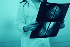
A mammogram is an x-ray picture of your breast. Mammograms are used to regularly check for breast cancer in women who may present with no signs or symptoms of the disease. Screening mammograms involve two x-ray pictures of each breast that are analysed by a radiographer for signs of abnormality. Free routine mammographic screening is available in each state for women aged 50-74 through BreastScreen Australia.*
*Women aged 40-49, or over 75, are also entitled to a free mammogram, however, they receive no reminder prompts, like women aged 50-74 do.
Book online or contact BreastScreen Australia on 13 20 50.
BIOPSY
A biopsy is the removal of a small sample of tissue from the breast or lymph nodes. The tissue is then examined by a pathologist (specialist doctor) under a microscope. This process helps to determine if the sampled tissue has any cancer cells. It also helps to determine the appropriate treatment plan.
MAGNETIC RESONANCE IMAGING (MRI)
An MRI produces an image of the inside of your body using magnetic fields. Women under 50 years of age who are at high risk of breast cancer are eligible for routine screenings with MRI through Medicare. To access this service, younger women must be referred by their GP or specialist.
ULTRASOUND
An ultrasound uses soundwaves to outline a part of your body. A breast ultrasound is used to see whether a lump found in the breast is solid or filled with fluid. An ultrasound is often used to check abnormal results from a mammogram.
What we’re researching now – harnessing the immune system to develop new ways of detecting and treating aggressive breast cancers
Problem: Most breast cancers express the estrogen receptor and are therefore classified as hormone responsive tumours. This means that they can be targeted with anti-estrogen treatment. Most patients respond very well to anti-estrogen therapy, but up to 20% of patients have a more aggressive form of this type of breast cancer where the treatment is not effective.
Solution: There is currently no way to predict which patients have this aggressive subtype, but A/Prof Blancafort’s has discovered a biomarker that may be useful for identifying these “hidden” subtypes of breast cancer. This project will confirm whether this biomarker can identify these hidden subtypes of estrogen receptor positive breast cancer and further investigate whether targeting this biomarker may result in a new therapeutic option for patients diagnosed with this disease.
DONATE NOW TO HELP FUND THIS RESEARCH
BREAST AWARENESS
It’s important to be familiar with your breasts so that you’re alert of any changes that might occur in the breast region. If you experience any symptoms such as lumps, dimples, discharge or discolouration, head to your doctor for further examination. It’s also important to be remember that finding a lump doesn’t mean that you have cancer; most lumps are benign (not cancer). So, don’t panic if you do find a change, make an appointment with your doctor for further evaluation.
